WattSearch
Your WattLeaderboard
Language
Autoplay narration
Appearance

Feedback

Have a question or an idea? Something not working? Send us a message.

What would you like to share?
Up to 3 files, 5 MB each, 12 MB total.
Or contact us directly

Direct contacts open a separate chat or email. Files attached here are not included.

arohalya@gmail.com
A little wonder, every day

How MRI took shape

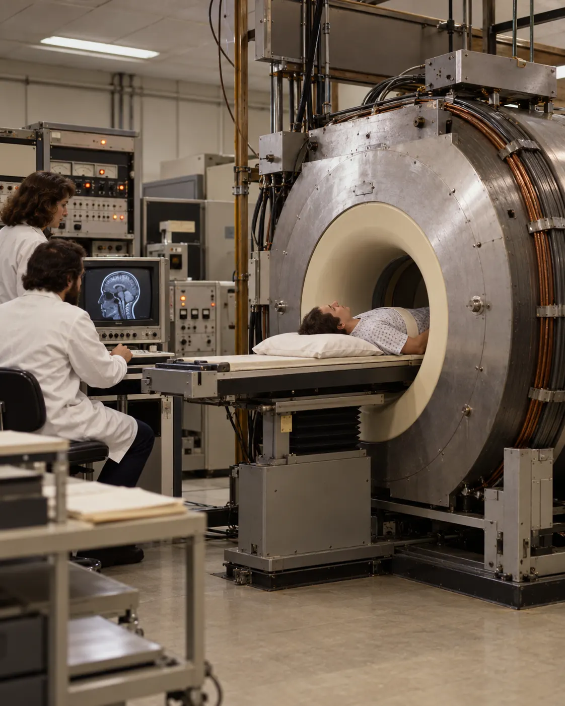
Титульна AI-концептуальна реконструкція ранньої дослідницької системи MR-зображення / AI conceptual reconstruction of an early experimental MR imaging setup.
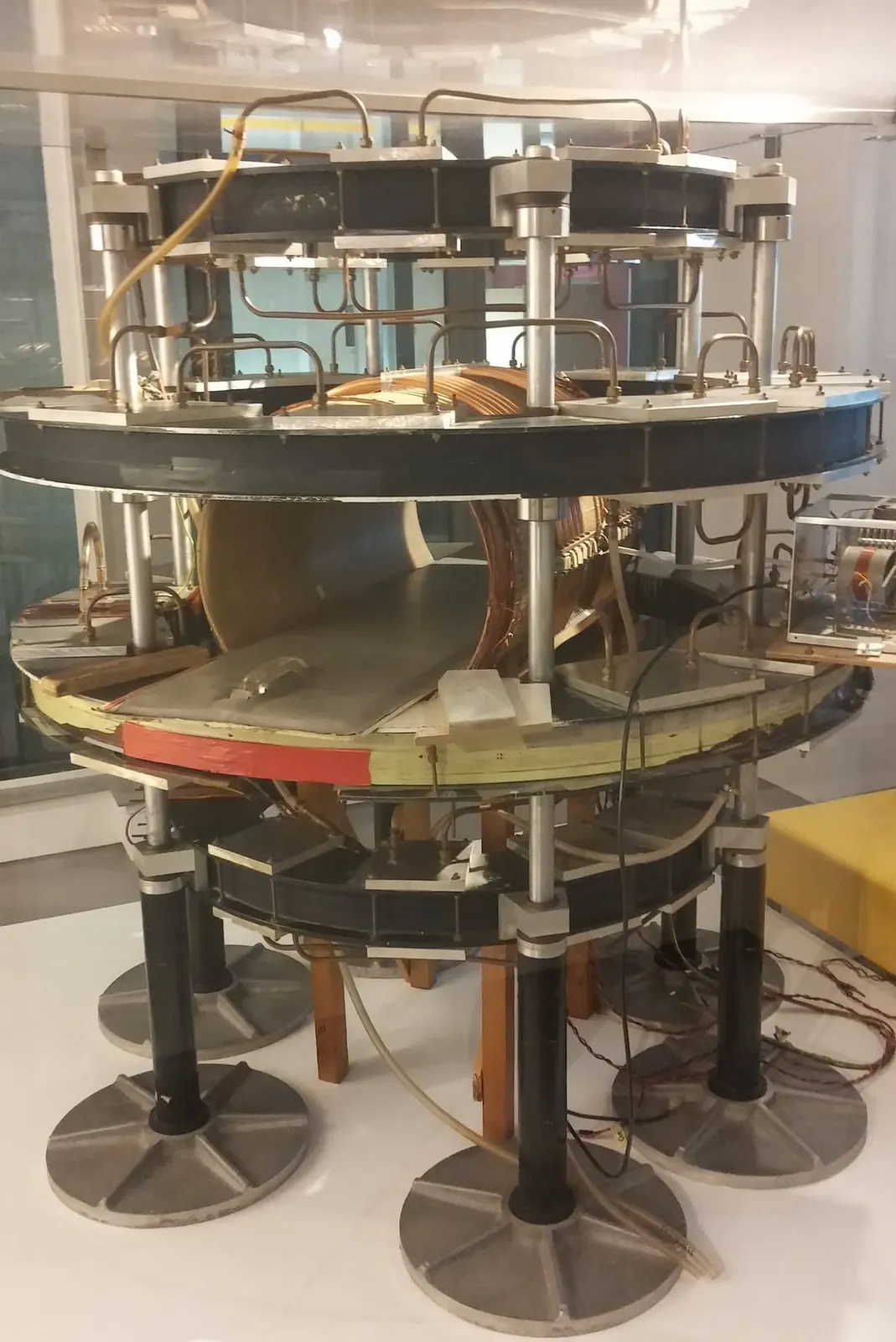
оптимізовано з `media/mark-one-mri-scanner.jpg`: 1170×1751 px (половина початкової ширини й висоти), JPEG quality 82.
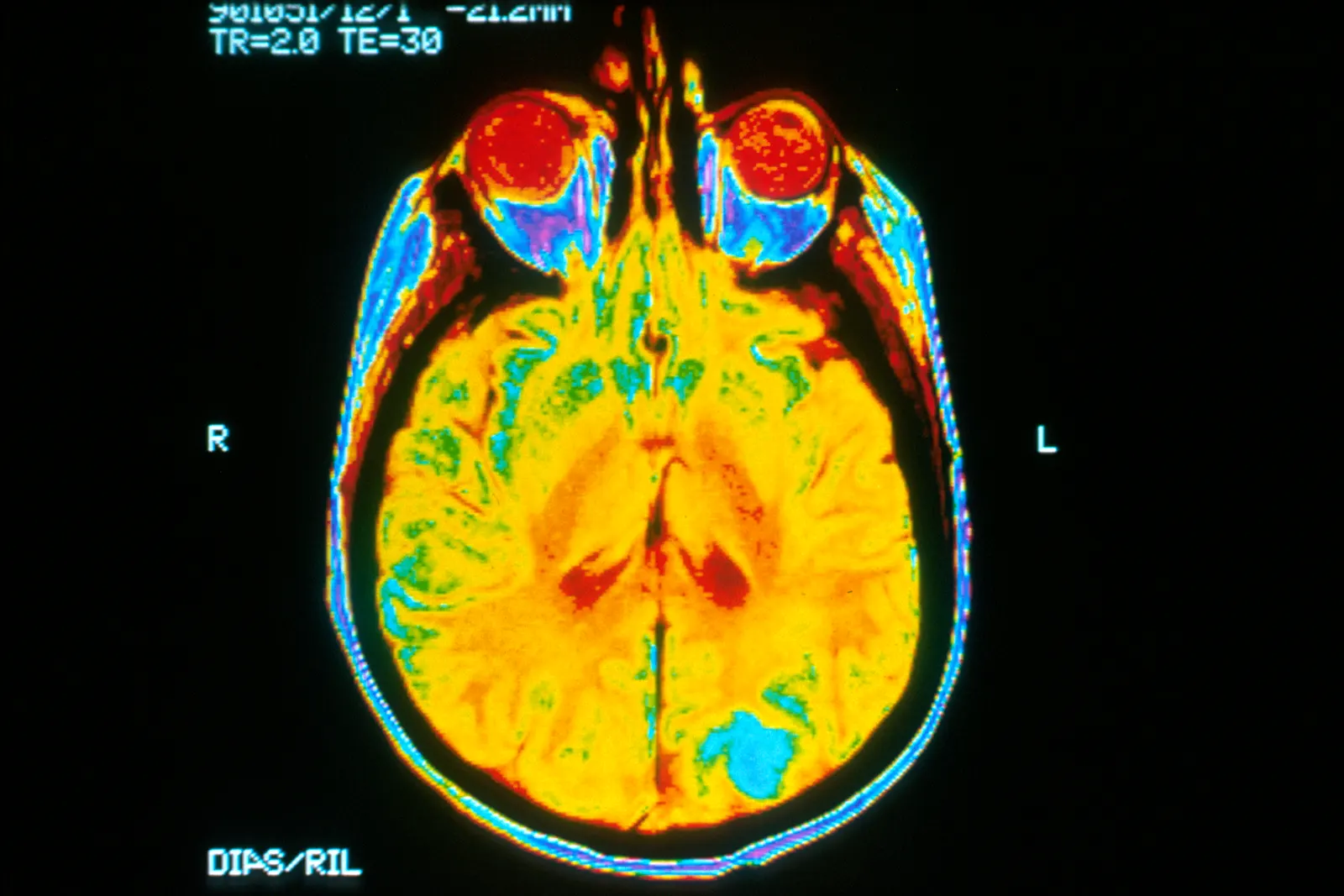
Документальне MRI-зображення головного мозку / documentary brain MRI image. Original page: https://commons.wikimedia.org/wiki/File:Brain_MRI.jpg. Source: Dr. Leon Kaufman, UCSF; released by the U.S. National Cancer Institute; photographer not identified. Public domain as a U.S. federal government image; credit NCI / UCSF. No edits. Modern/contextual result, not a 1977 scan.

Титульна AI-концептуальна реконструкція ранньої дослідницької системи MR-зображення / AI conceptual reconstruction of an early experimental MR imaging setup.

Listen to fact0:00 / —

MRI took shape through several related contributions. In 1973, Paul Lauterbur published a method for producing spatial images with nuclear magnetic resonance; Peter Mansfield developed mathematical and technical methods that made such imaging more practical. The 2003 Nobel Prize in Physiology or Medicine was awarded jointly to them for discoveries concerning magnetic resonance imaging. Separately, in 1977 Raymond Damadian, Michael Goldsmith, and Lawrence Minkoff published an account of the first chemical image of a living human: a cross-section of the chest produced with their FONAR system.